

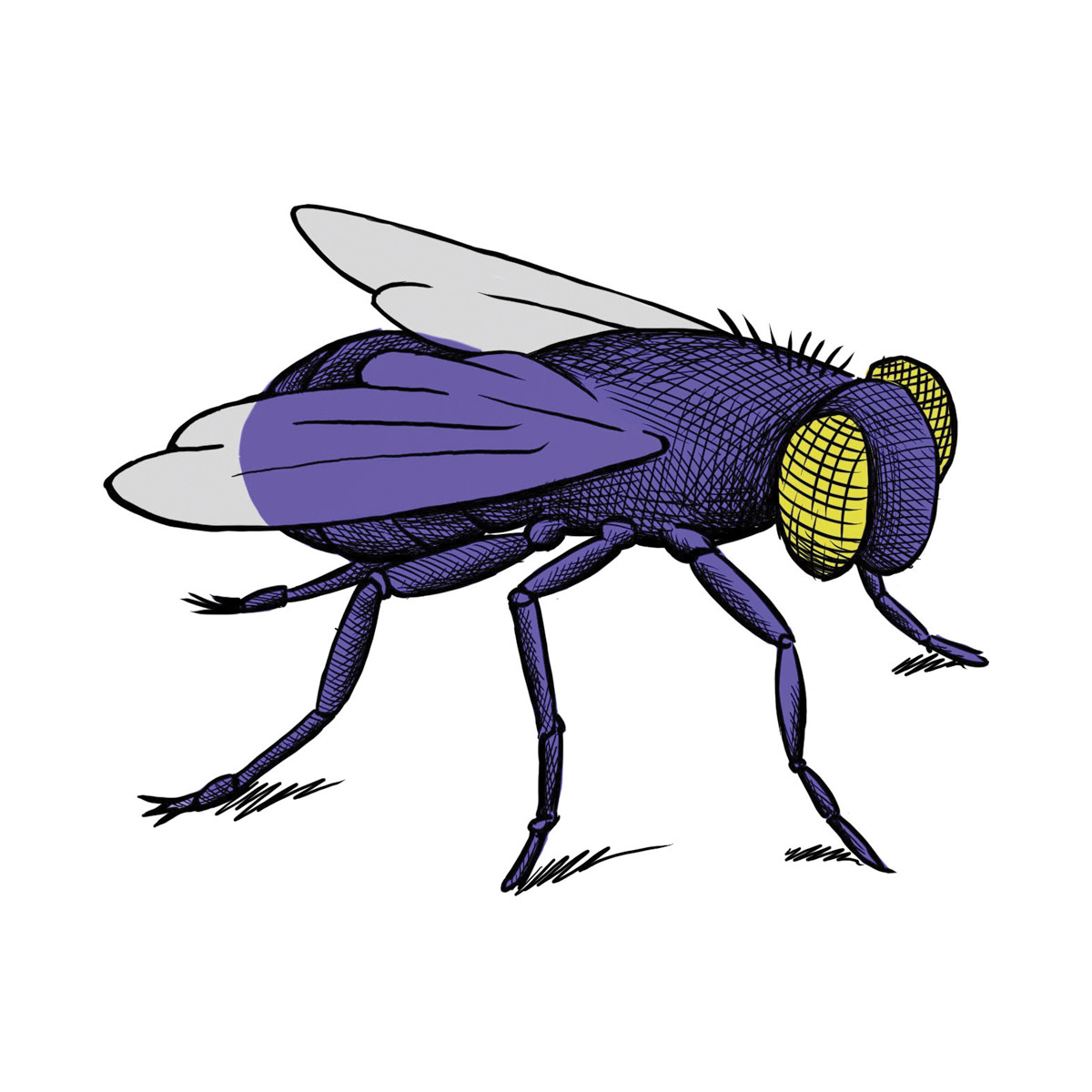
See hairs on the leg of a fly, spores on a fern, and nanowires that are a hundred thousand times thinner than the diameter of a human hair — researchers at Colgate now have this ability with a new field emission scanning electron microscope (FE-SEM).
Funding for the FE-SEM came from a $439,805 National Science Foundation (NSF) grant. Complete with a suite of high-tech detectors, the FE-SEM will aid faculty and also train students for advanced research in geology, physics, biology, computer science, and other fields.
“[The detectors] are incredibly versatile,” says primary investigator (PI) Martin Wong, professor of earth and environmental geosciences. “So many different things happen when electrons hit a surface, and you can use all those different signals to examine the composition and structure of your sample.” With a much smaller wavelength than visible light, electrons allow researchers to image vastly tinier objects than is possible with an optical microscope.
Here are some of the applications:
→ Wong’s research focuses on plate tectonics of how mountain belts form and continents split.
“When rocks are hot and deep, they don’t snap or break, but they actually flow like silly putty or saltwater taffy,” says Wong, who focuses on an area of the American West called the Basin and Range Province. He analyzes samples with a special detector called an electron backscatter diffraction detector, which is able to show how crystals are oriented within it. “That tells us a lot about which directions they were stretching in, the temperatures at which they were doing that, and how deep inside the Earth they were.” While Wong is mostly interested in the basic science behind such processes, understanding them can also aid in earthquake detection and location of rare minerals.
→ Geology professor William Peck studies the formation of rocks in the Adirondack Mountains, Ontario, and New Jersey.
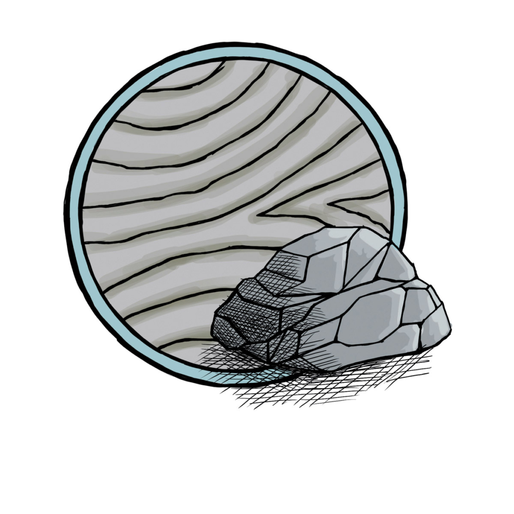
He’ll be able to use the electron microscope to image samples with something called the secondary electron detector, which is able to display a graded map that differentiates between minerals. For a more in-depth analysis, he’ll use an X-ray energy spectrometer, which can measure the degree that the electron stream excites the atoms within a sample, emitting X-rays that provide a fingerprint of which specific elements are contained within.
→ Ramesh Adhikari, assistant professor of physics, works with organic materials.
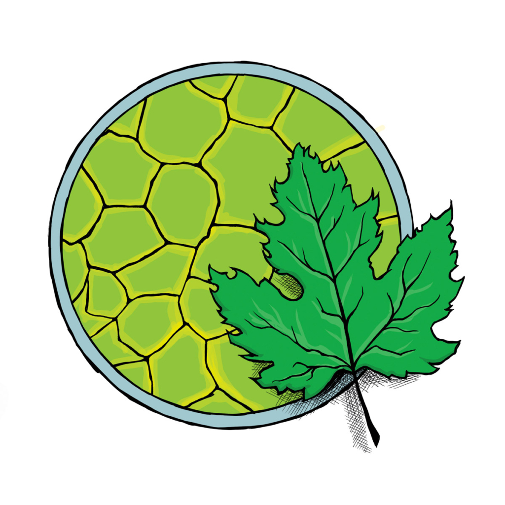
One project, for example, coaxes amino acids to self-assemble into tiny nanotubes; another threads nanoscopic computing components into the veins of leaves. “These bio-based materials tend to absorb the electrons that fall onto them, so you don’t really see that much,” Adhikari says. That problem can be fixed by turning up the voltage of the beam; however, that actually damages organic samples. The new microscope, however, can produce high-resolution images at a very low voltage without damaging fragile organic components, allowing Adhikari to examine the tiny structures he is creating. Among other uses, he is embedding the nanotubes into a polyester fabric to create a hydrophobic material that can filter oil from water. The leaves can be used to create biodegradable electronic equipment.
→ The ability to create crisp images at a low voltage is also essential for the work of Rebecca Metzler, professor of physics, who studies biomineralization, the process by which marine organisms create shells and other hard materials.
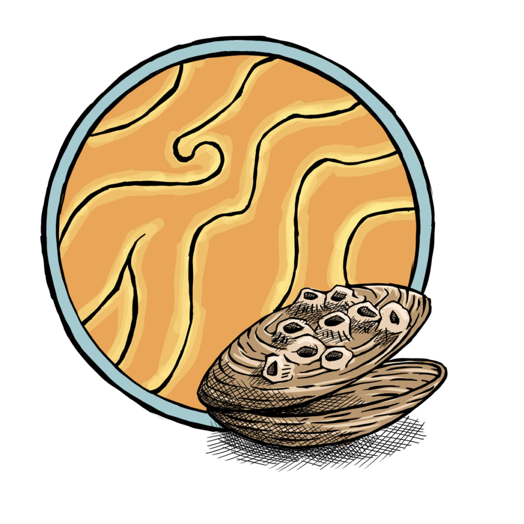
Some of her work examines exoskeleton formation by juvenile barnacles, which can be a scant 100 microns wide. “Our current scanning microscope can’t resolve the crystals making up their exoskeletons,” says Metzler, who has previously had to make the 75-mile trip to Cornell to use its more advanced equipment. She also studies other species of clams from the Gulf of Mexico, using the X-ray spectrometer to identify elements, and the backscatter diffraction detector to examine how crystals are oriented within shells in order to see how climate change affects the durability of shells over time.
In addition to these projects, the microscope will be used by a variety of faculty, studying everything from volcanic eruptions in the Galápagos Islands to wear patterns of tools at pre-Hispanic archaeological sites. Countless students will use it for theses projects over the next couple of decades; at the same time, exposure to the advanced instrument will provide students with experiences that could help them in working with microprocessors, nanotechnology, or mining. “This is a broadly used piece of equipment with all sorts of research and industry applications,” Wong says. “Being trained on it will give students a leg up no matter what avenue they pursue.”